Search
Optic neuritis.

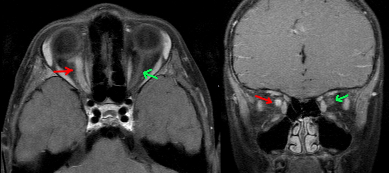
5 year old patient with 1 week of right eye blurry vision, then several days of right eye pain. Physical exam notable for right papilledema and progressively worsening vision.
MRI postcontrast through the orbits shows an enlarged, hyperenhancing right optic nerve (red arrow) compared to the normal left side (green arrow), compatible with optic neuritis.
A lumbar puncture was performed: no oligoclonal bands, no aquaporin 4 IgG, positive anti-MOG. The patient was treated with prednisone with return to normal vision a few months later.
Final diagnosis: optic neuritis from myelin oligodendrocyte glycoprotein antibody-associated disease (MOGAD).
12-ft fall with loss of hearing.

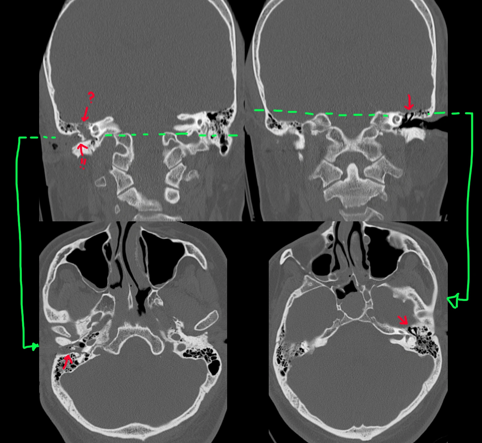
31 year old doing some doing roof work when he fell. Seen in the ED with right ear bleeding and right-sided decreased hearing.
The normal left side is depicted in the right images (standard convention). Coronal view [top right] shows the middle ear ossicles (arrow) in the middle ear cavity, and axial view [bottom right] shows the normal "ice cream cone" appearance (arrow).
By comparison, the right middle ear ossicles are missing in the middle ear cavity [top left] (arrow with ?) and found in the external auditory canal (arrow with !!) instead. Axial view [bottom left] shows a distinct lack of the ice cream cone appearance. The middle ear and external ear canals are also filled with fluid/blood. The tympanic membrane is clearly ruptured since the middle ear ossicles are outside.
Nasal proboscis.
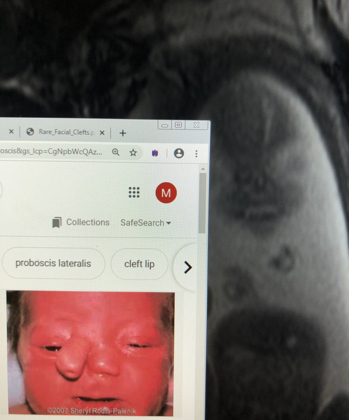
Not the best quality, but this is the only image I saved of this fetal MRI, with a random internet picture of what it would look like after birth.
Gnathic (jaw) osteosarcoma.

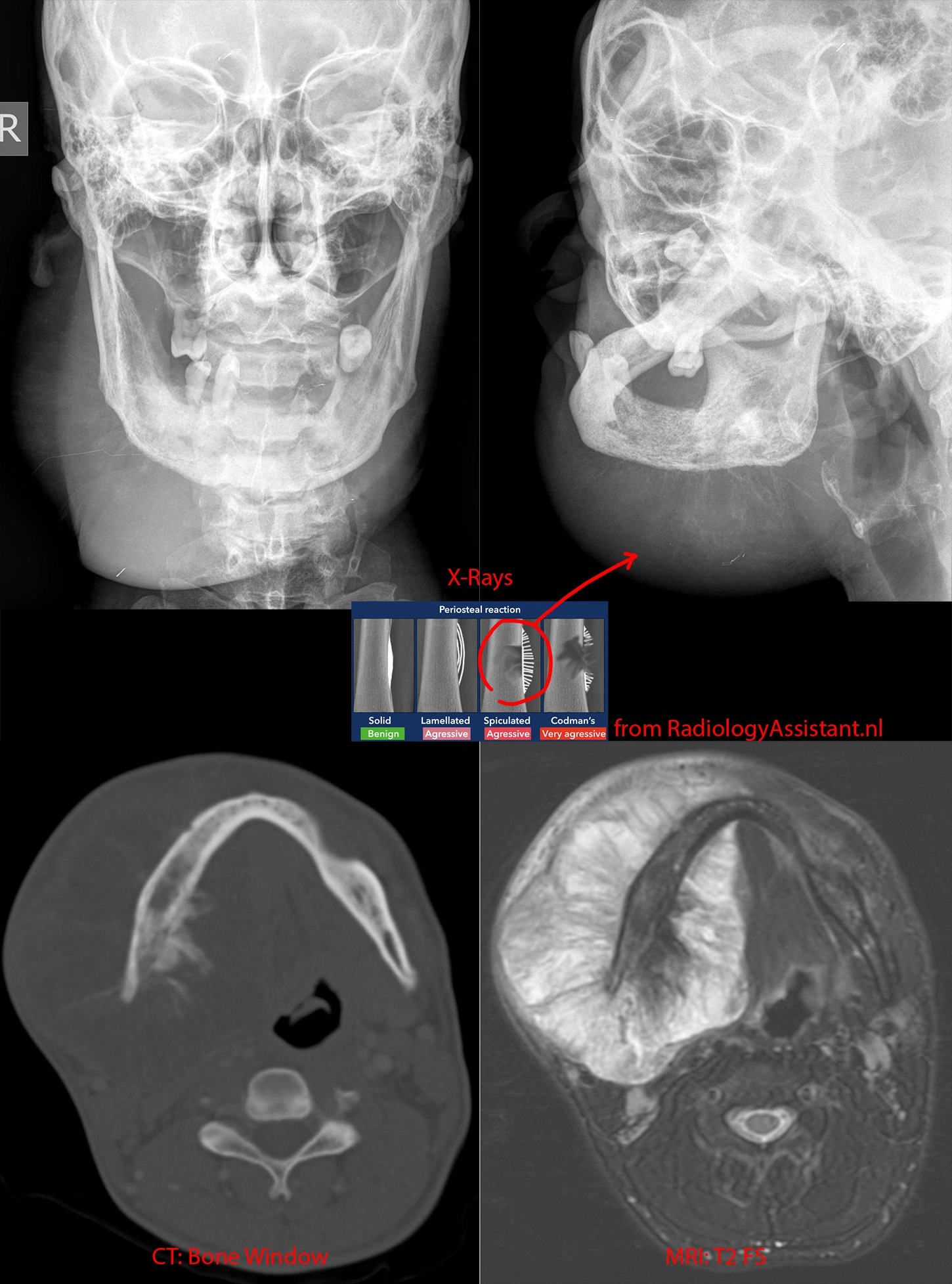
Top left and right: X-rays showing a BIG right jaw mass. Whispy calcifications likely represent spiculated periosteal reaction, a very aggressive bony reaction.
Bottom left: CT in the bone/calcification window shows the periosteal reaction better.
Bottom right: MR in the T2 sequence shows the full extent of the jaw mass and how it extends far beyond the mandible's margins, both outward and inward into the floor of mouth.
While this mass is very large, in general, jaw osteosarcoma has a better prognosis than conventional osteosarcomas.
Traumatic globe rupture.


Past medical history of seizure. Had an unwitnessed fall.
CT shows right globe with abnormal irregular morphology, abnormal location/appearance of the lens, and presence of air within the globe. On the coronal view, there is fluid infiltrating the surrounding orbital fat. A left forehead hematoma is also present.
Fibrous dysplasia.

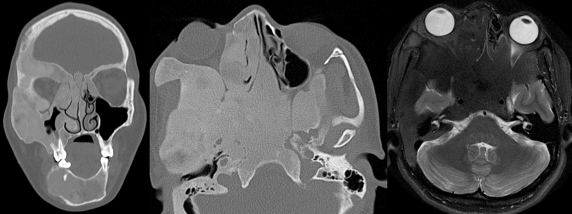
Female in her 20s with (all right-sided) proptosis, intermittent vision loss, facial pain, and mid to lower face numbness.
Coronal and axial CT images show a homogenous bony expansile process that results in narrowing of the soft tissue compartments of the face. As a result, the right eye is pushed forward (proptosis; as seen on the MR image), and many of the right-sided (as well as some left-sided) skull base foramina that carry nerve bundles and blood vessels are severely narrowed.