Brain Abscess. [Neuroradiology] [CT] [MR]
Brain Abscess. [Neuroradiology] [CT] [MR]


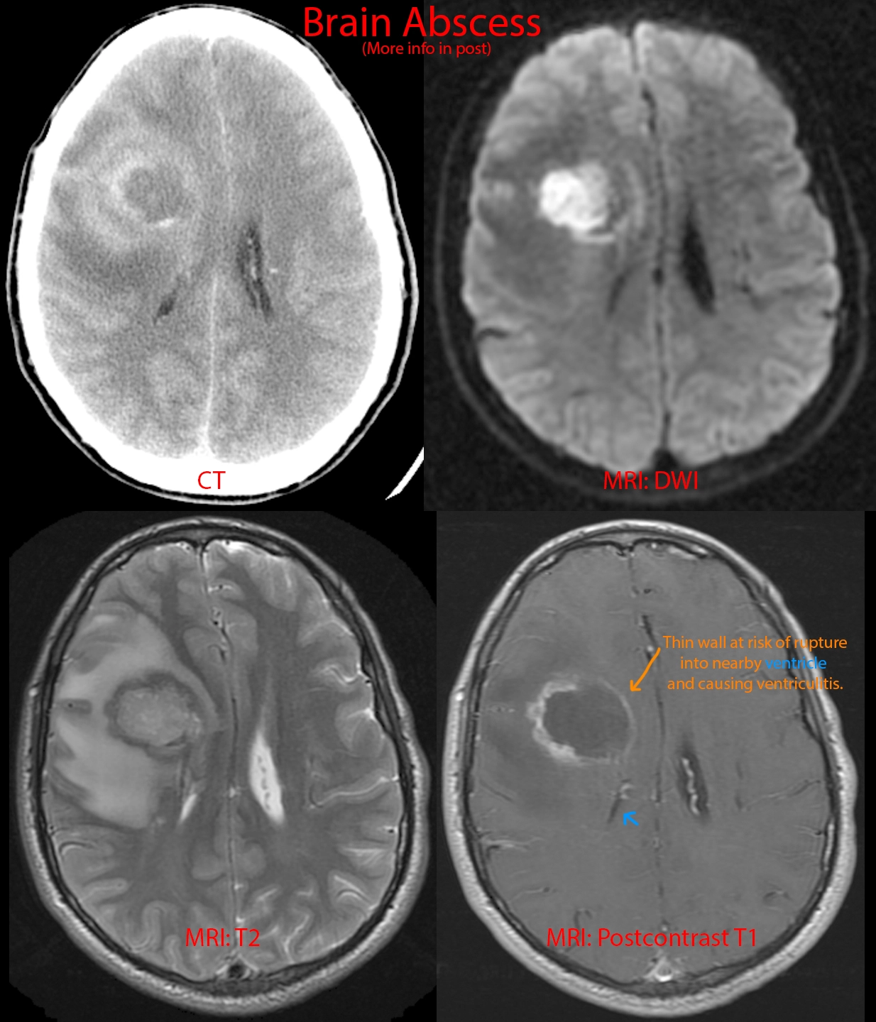
28 year old male with a history of complex congenital heart disease presenting with altered mental status.
CT [top right image]: 4 cm centrally necrotic mass and prominent surrounding brain edema. Ddx: abscess versus tumor.
DWI [top left image]: True diffusion restriction within the central part of the mass, suggesting pus.
T2 [bottom right image]: Great example of the bright-dark-bright arrangement of abscesses on the T2 sequence (bright center, dark abscess capsule, bright surrounding edema).
Postcontrast T1 [bottom left image]: The abscess capsule appears as rim enhancement. The thin medial wall of the abscess [orange arrow] is growing every closer towards the adjacent lateral ventricle [blue arrow] and will ultimately rupture into the ventricle, causing ventriculitis, a high mortality situation.
This patient required surgical drainage of the abscess. The bacteria responsible was a Streptococcus species. Congenital heart disease is a risk factor for cerebral abscesses.