Search
Ovarian mucinous cystadenocarcinoma.

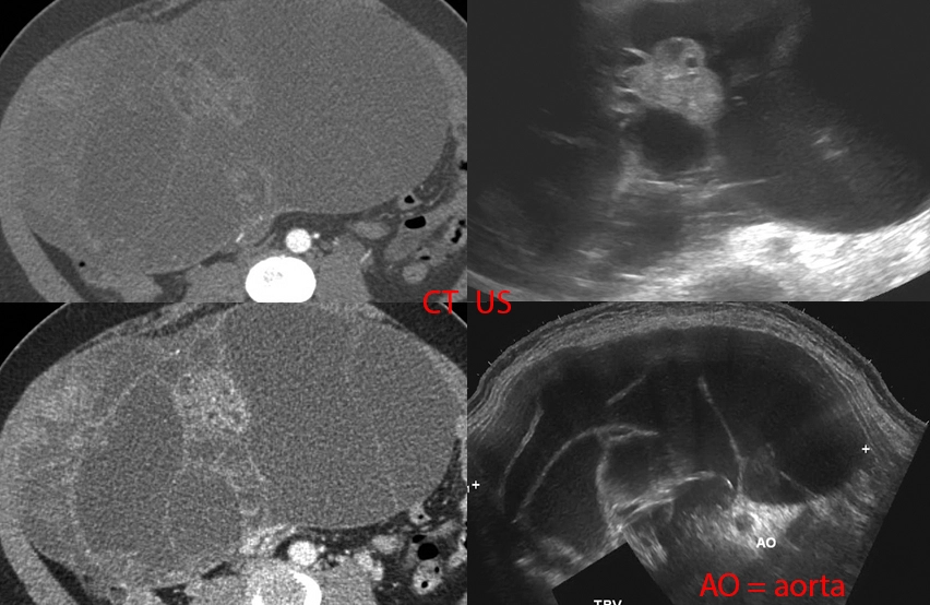
33 year old female with abdominal pain, abdominal distention, nausea/vomiting, early satiety, and weight loss.
Bottom right: Ultrasound done in a panorama shows how distended the abdomen is by a large multi-cystic mass.
Top right: Non-panoramic ultrasound image shows how limited the imaging modality is in being able to cover such a large mass. This image also shows a more solid area within the mass.
Left: CT images approximately where the ultrasound was done.
The patient underwent laparotomy with removal of the ovarian, fallopian tube, and appendix. There was a large ovarian cyst that was draining serous fluid (watery), mucinous fuid (mucus-like), and blood. The final path was as titled.
Right kidney stone.


25 year old with right flank and pelvic pain for 2 days. No fevers, chills, nausea, vomiting, or other urinary symptoms however.
[Top]: Ultrasound shows right kidney with distended pelvis (hydronephrosis).
[Mid & Bottom]: Ultrasound transverse and sagittal images shows a 4.5 mm stone at the very end of the ureter (ureterovesicular junction).
Pretty straightforward case. For the nonmedical visitors, this is what we look for if your doc wants to get an US for flank pain / kidney stone suspicion. See the other case for a CT version.
Acrania-Exencephaly-Anencephaly Sequence.


Mom was a 36 year old with active meth abuse, homelessness, and scant prenatal care.
Prenatal ultrasound at 36 weeks shows a highly deformed fetal brain with large fluid-filled spaces that might represent cysts or hydrocephalus. The brain contours are lobulated and irregular due to absence of an overlying skull.
She was recommended for pregnancy termination but no-showed to that procedure. She then presented 3 weeks later, at 39 weeks, in labor. The baby was delivered spontaneously. The baby had absent skull above the level of the ears. The brain was large and could be seen through a membranous sac. The baby passed away one day later.
Acrania is the absence of a skull. Exencephaly is the condition where a brain (which may have otherwise been normal) is not covered by the skull in the prenatal setting. Due to the exposure of brain tissue to amniotic fluid, which is toxic to brain, exencephaly will invariably lead to anencephaly, which is the absence of all/most of the brain.
Thoracic myelomeningocele, dermal sinus tract, dermoid.


Otherwise healthy newborn with "abnormal back finding."
Ultrasound shows, underneath the back "mass," a defect of the posterior spine through which the spinal sac protrudes.
MRI confirms the defect and spinal sac protrusion. Additionally, there was some tissue that was thought to represent neural placode within the herniated sac. There was no obvious fat-containing mass within to suggest dermoid by imaging.
Surgery showed absent T6-T7 spinal lamina with a fibrous tract extending from the back of the spinal cord to the skin surface. Hair and other skin-related debris extruded from the tract as it was opened. The entire fibrous tract and its contents were removed.
The classic internet rectal foreign body... or is it?
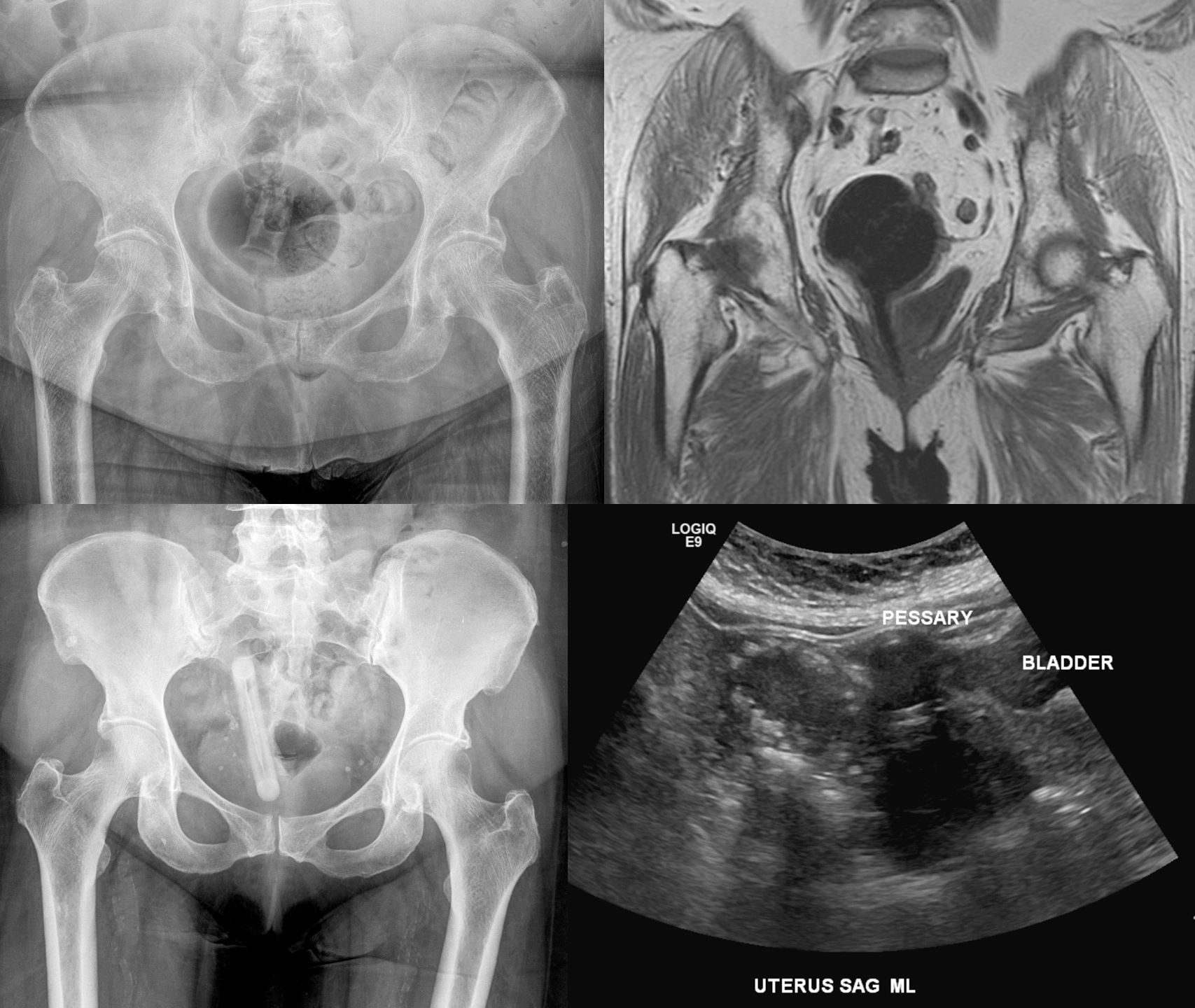
On the left side, 2 x-rays of 2 different patients with 2 different objects in their pelvis. Internet denizens would no doubt instantly diagnose these as accidental rectal foreign bodies from people falling on their butts. However, these are both pessaries used to treat pelvic organ prolapse. The top one is an inflatoball pessary. The bottom one is a ring-shaped pessary that we see on edge.
Leriche syndrome (aortoiliac occlusive disease).

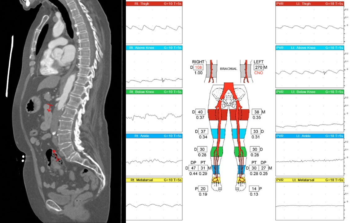
62 year old female with a history of tobacco, opioid, and meth abuse as well as heart attack requiring coronary bypass and stents. She presents with bilateral lower extremity claudication (painful fatigue with physical activity) starting from her glutes.
CT angiogram shows occlusion of the lower aorta (red markings) as well as both common iliac arteries.
Vascular ultrasound shows very poor arterial waveforms throughout the lower extremities, essentially flat/absent below the knee.
She underwent a transabdominal aortobifemoral bypass to treat this.
Radiopedia article on Leriche syndrome.

