Search
Painful left shoulder.

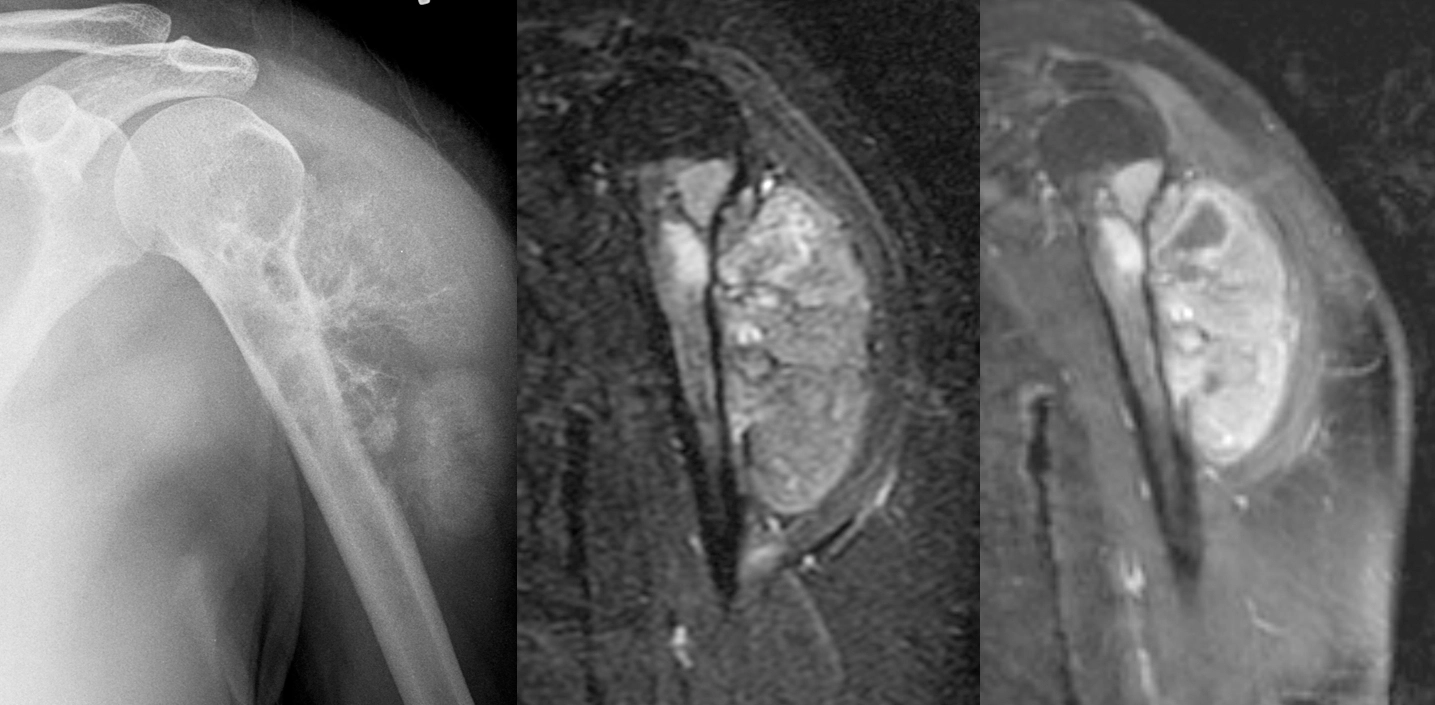
Female in her 30s with painful left shoulder.
[Left]: X-ray shows a mass arising from the left proximal humerus and extending into the adjacent shoulder soft tissues with really aggressive periosteal reaction ("hair on end"). The proximal humerus itself is also heterogeneous with lucent areas. The lateral surface of the upper humerus shows "saucerization," where the cortex is thinned out and looks like a saucer seen on edge.
[Middle]: MRI IR sequence shows a hyperintense bony mass with large soft tissue component.
[Right]: MRI postcontrast T1 IDEAL shows that the mass is enhancing.
This turned out to be high-grade surface osteosarcoma.
10 year old with insidious onset of right medial thigh pain.
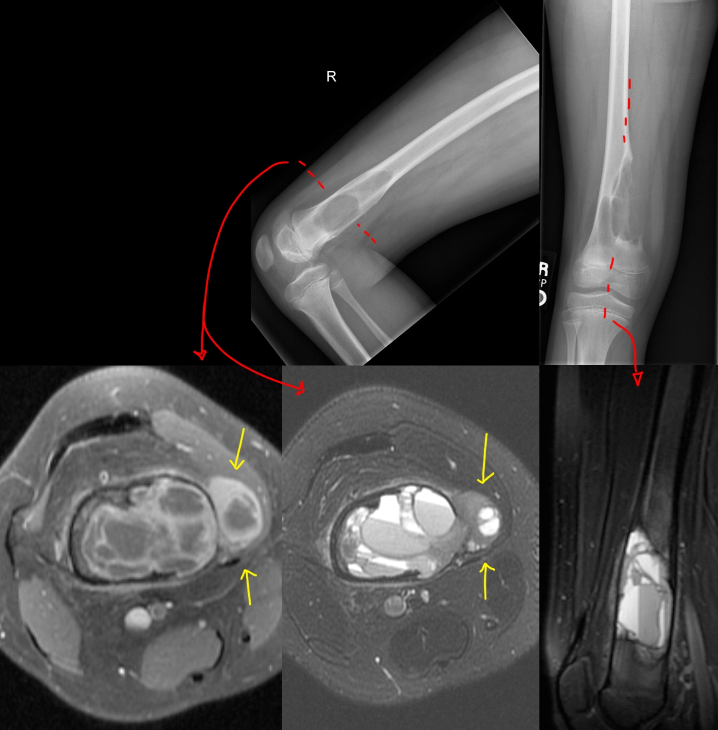
[Top]: X-ray shows a lucent, bubbly, lesion of the distal femur at the metaphysis. On the frontal view [top right], there is breakage through the medial femoral cortex into the adjacent soft tissues, not a good sign.
[Bottom]: MRI shows a multicystic lesion filling the distal femur containing multiple locules, many with fluid-fluid, fluid-debris, and fluid-hemorrhage levels. The most common lesions with this striking appearance are aneurysmal bone cyst, giant cell tumor, or telangiectatic osteosarcoma. Unfortunately, there is clearly extension of the bone tumor beyond the bone (yellow arrows), which favors a more aggressive neoplasm from that differential diagnosis - this turned out to be telangiectatic osteosarcoma.
5 year old who fell off a slide with an unexpected finding.


5 year old who fell off a slide.
Initial imaging shows a comminuted fracture through the distal humerus, compatible with a supracondylar fracture. Nothing else appreciable here, except maybe in retrospect some lucency of the distal humerus where the fracture is.
4- and 7-month follow-up radiographs shows a growing lucent lesion of the distal humerus, expanding the bone there. It has a multicystic appearance. A diagnosis of large simple bone cyst versus aneurysmal bone cyst was proposed.
12 month follow-up was done after the cyst was opened surgically, its contents scraped off, and the resulting cavity was packed with allograft bone chips. At surgery, this turned out to be an aneurysmal bone cyst.
5 year follow-up shows involution of the cyst cavity with some residual heterogeneity and a bone spur at the anterior aspect of the distal humerus.
Gnathic (jaw) osteosarcoma.

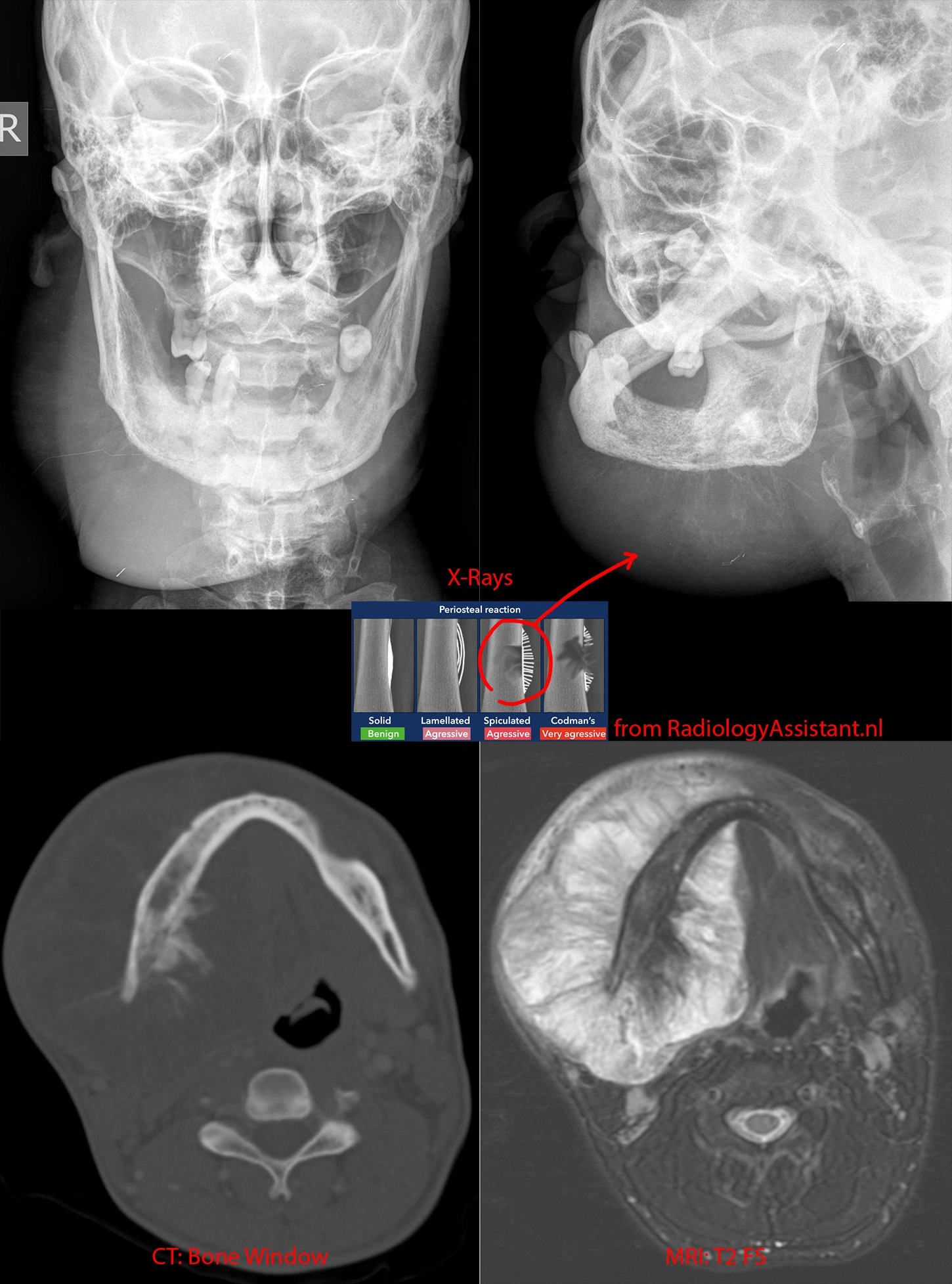
Top left and right: X-rays showing a BIG right jaw mass. Whispy calcifications likely represent spiculated periosteal reaction, a very aggressive bony reaction.
Bottom left: CT in the bone/calcification window shows the periosteal reaction better.
Bottom right: MR in the T2 sequence shows the full extent of the jaw mass and how it extends far beyond the mandible's margins, both outward and inward into the floor of mouth.
While this mass is very large, in general, jaw osteosarcoma has a better prognosis than conventional osteosarcomas.
Innumerable skull lesions of multiple myeloma.

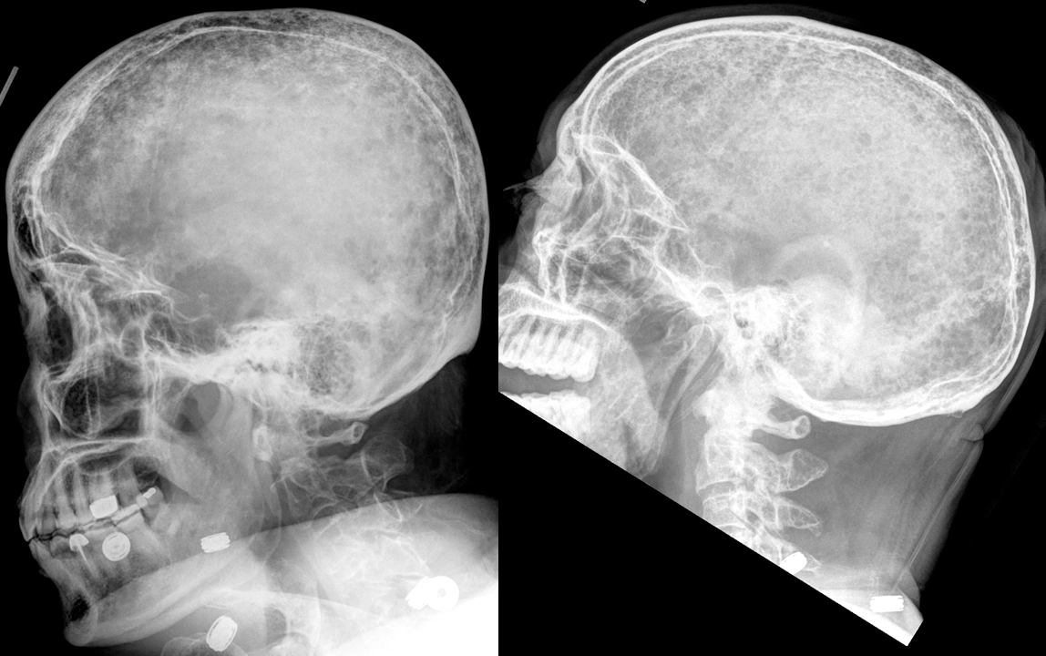
Two different patients, both aged 59, with known multiple myeloma.
Codman's triangles in osteosarcoma.

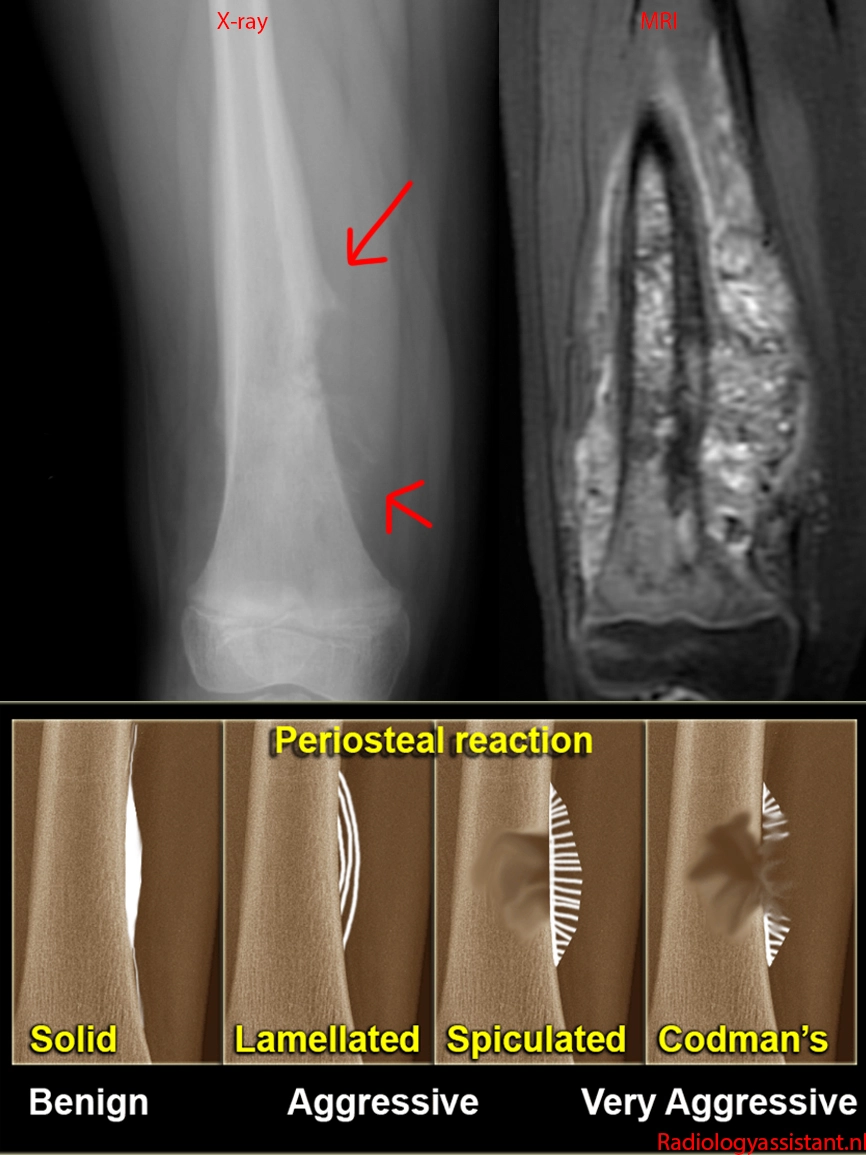
7 year old with conventional (intramedullary high-grade) osteosarcoma of the femur.
X-ray shows an aggressive bone lesion with Codman's triangles and extension into the adjacent soft tissues.
MRI shows the full extent of the tumor much better - it is involving the full thickness of the femur, has much further soft tissue extension than suggested by the Codman's triangles, and even extends into the soft tissues on the other side.
Diagnosis - Lululemon.

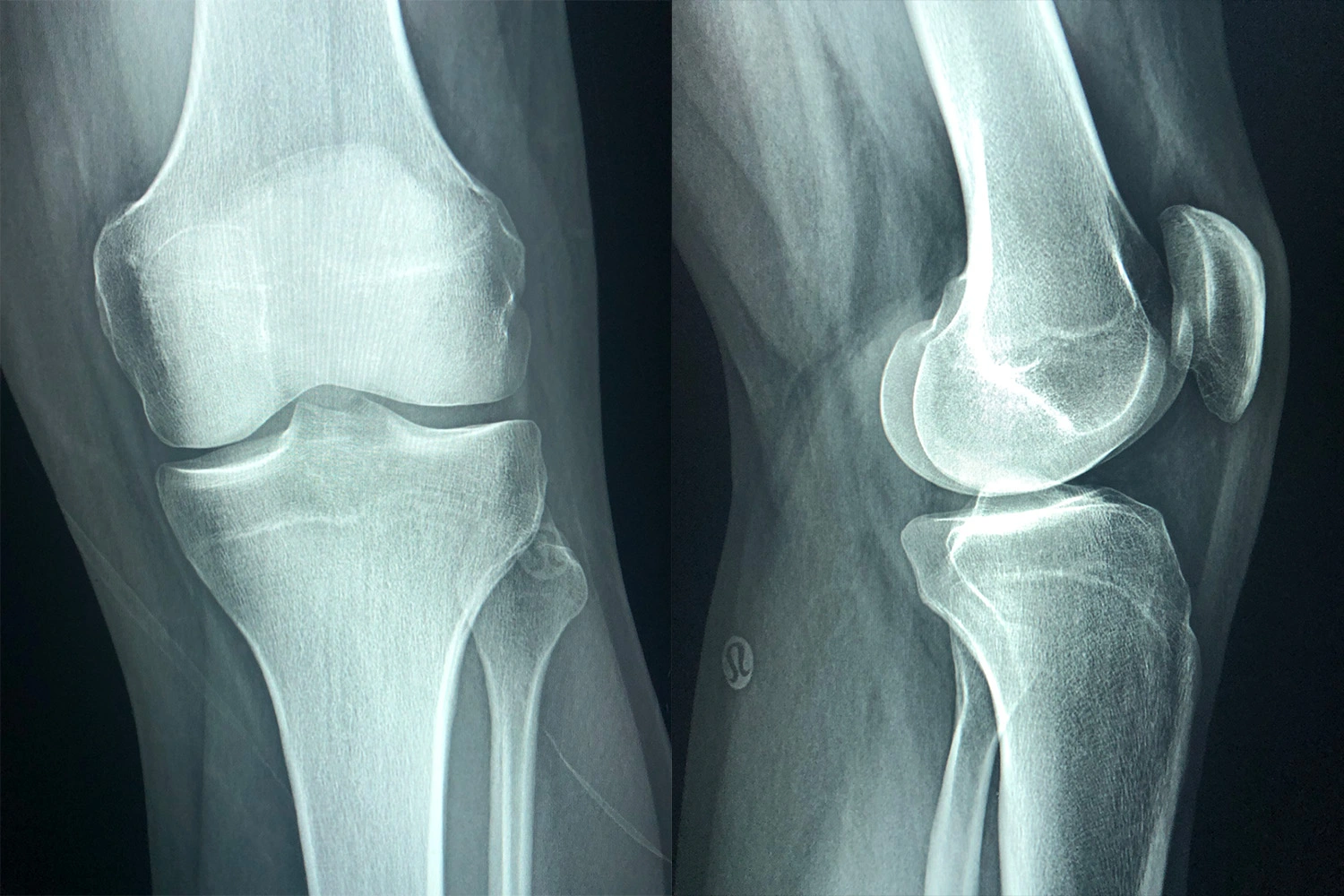
If you're planning on getting an MRI, avoid wearing Lululemon, especially their silver-impregnated stuff - the strong changing magnetic fields can cause the clothing to heat up to the point of causing burns (just Google for news articles).
Life's middle finger: malignant melanoma.

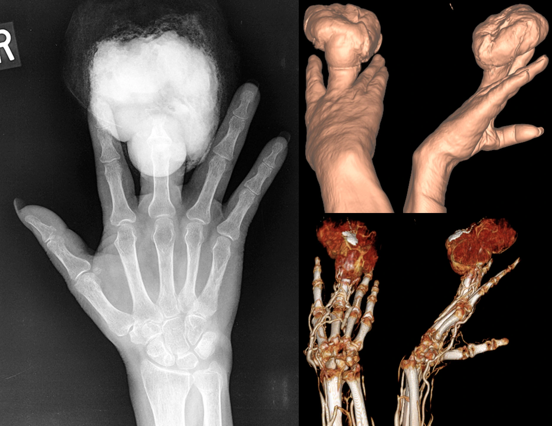
54 year old female with a history of minor injury to the finger 6 months before these studies were obtained, subsequently developed an infection requiring debridement. The wound was then stable until 2 months before, when a large, fungating, hemorrhagic mass grew.
X-ray shows large radiopaque mass eroding the distal finger (distal phalanx and distal portion of the middle phalanx).
CT 3D surface reconstructions show the morphology of the mass.
CTA 3D reconstruction shows the mass is very hypervascular.
Lymph node scintigraphy was performed showing a sentinel node at the axilla (not shown). The patient underwent amputation of the finger with axillary sentinel node biopsy, which was positive for metastatic melanoma. The patient was then lost to follow-up.
Accidentally fired gun into knee.

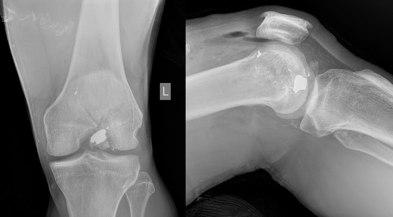
38 year old guy fired his 45 mm into his knee while cleaning the gun. No exit wound.
There is a comminuted fracture of the distal femur with multiple bullet fragments, including the largest at the intercondylar notch. There is gas/air in the knee joint, forming air-fluid levels on lateral view. There are additional foci of gas in the surrounding soft tissues.
Patient underwent arthrotomy for foreign body removal and fixation of the supracondylar/intercondylar femoral fracture.

