Search
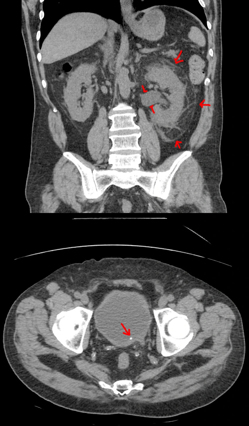
Left kidney stone.


57 year old with left flank pain for 12 hours. Urine sample 2+ for blood. White count at 14.
[Top]: Coronal CT shows the left kidney is enlarged, with angry-looking, inflamed, surrounding fat. The renal pelvis is dilated (hydronephrosis).
[Bottom]: Axial CT shows a small stone at the very end of the ureter (ureterovesicular junction).
Pretty straightforward case. For the nonmedical visitors, this is what we look for if your doc wants to get a CT for flank pain / kidney stone suspicion. See the other case for an ultrasound version.
Right kidney stone.


25 year old with right flank and pelvic pain for 2 days. No fevers, chills, nausea, vomiting, or other urinary symptoms however.
[Top]: Ultrasound shows right kidney with distended pelvis (hydronephrosis).
[Mid & Bottom]: Ultrasound transverse and sagittal images shows a 4.5 mm stone at the very end of the ureter (ureterovesicular junction).
Pretty straightforward case. For the nonmedical visitors, this is what we look for if your doc wants to get an US for flank pain / kidney stone suspicion. See the other case for a CT version.
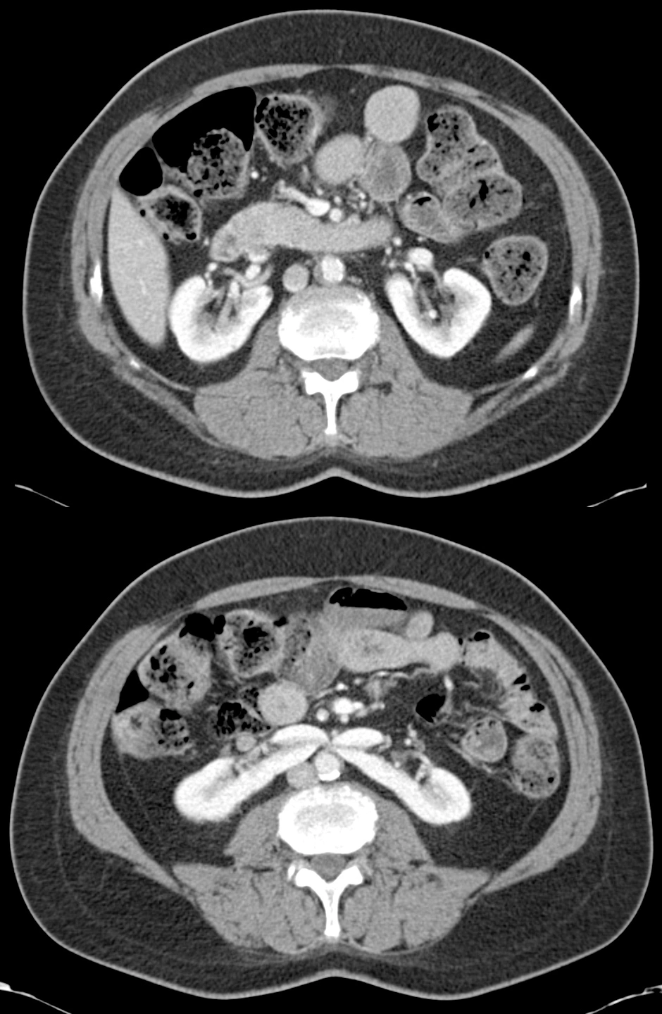
Horseshoe kidney.


CT obtained for some unrelated symptoms with an incidental finding.
Near the top, the two kidneys looked normal* [top image]. Near the bottom, the two kidneys wanted to join up into one kidney [bottom image].
*Technically, a little bit of renal malrotation due to the abnormal positioning from the horseshoe morphology.
This is generally a benign finding, but the abnormal shape might contribute to poor urinary drainage, which can cause renal stones, urinary infection, increased risk for cancer from the chronic inflammation caused by repeated infections, or increased risk of injury from trauma.
Metastatic renal cell carcinoma.


61 year old male, who came in with symptoms of right arm weakness and 3 months of weight loss despite trying to gain weight.
Head CT shows multiple round lesions in the (patient's) left cerebral hemisphere (right side on images), which likely account for the right-sided weakness. These lesions show hemorrhage as well as substantial surrounding brain edema.
A work-up for metastatic disease included a CT of the abdomen/pelvis, which showed the primary mass in the right kidney.