Search
Cancer versus not cancer on esophagram.

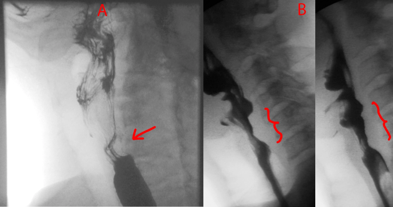
Quick one today. Take a look at Patient A and Patient B.
Patient A has a smooth focal indentation of the posterior cervical esophagus.
Patient B has a broader indentation that is also irregular and nodular along its contour.
Patient A has a cricopharyngeal bar, which is a prominence caused by the cricopharyngeus muscle that can cause dysphagia if it gets really prominent. Patient B has esophageal squamous cell carcinoma.
Small bowel obstruction due to gallstone ileus.
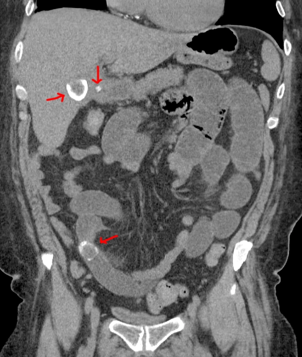
Red arrows point to 2 big gallstones, top one in the gallbladder and bottom one obstructing a small bowel loop, and a small gallstone in the cystic duct.
Small bowel obstruction from incarcerated inguinal hernia.

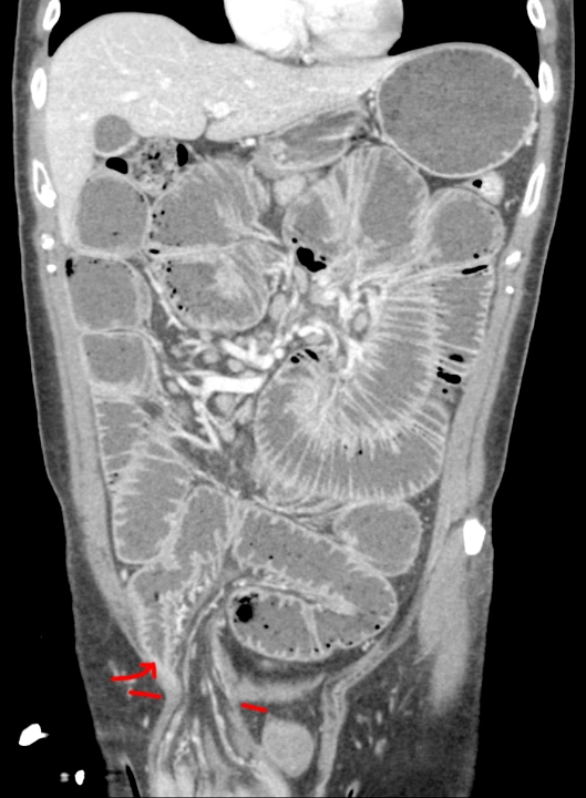
Red lines point to hernia entry. Red arrow points to where the bowel tapers and becomes obstructed as it enters the hernia sac.
Crohn's disease.

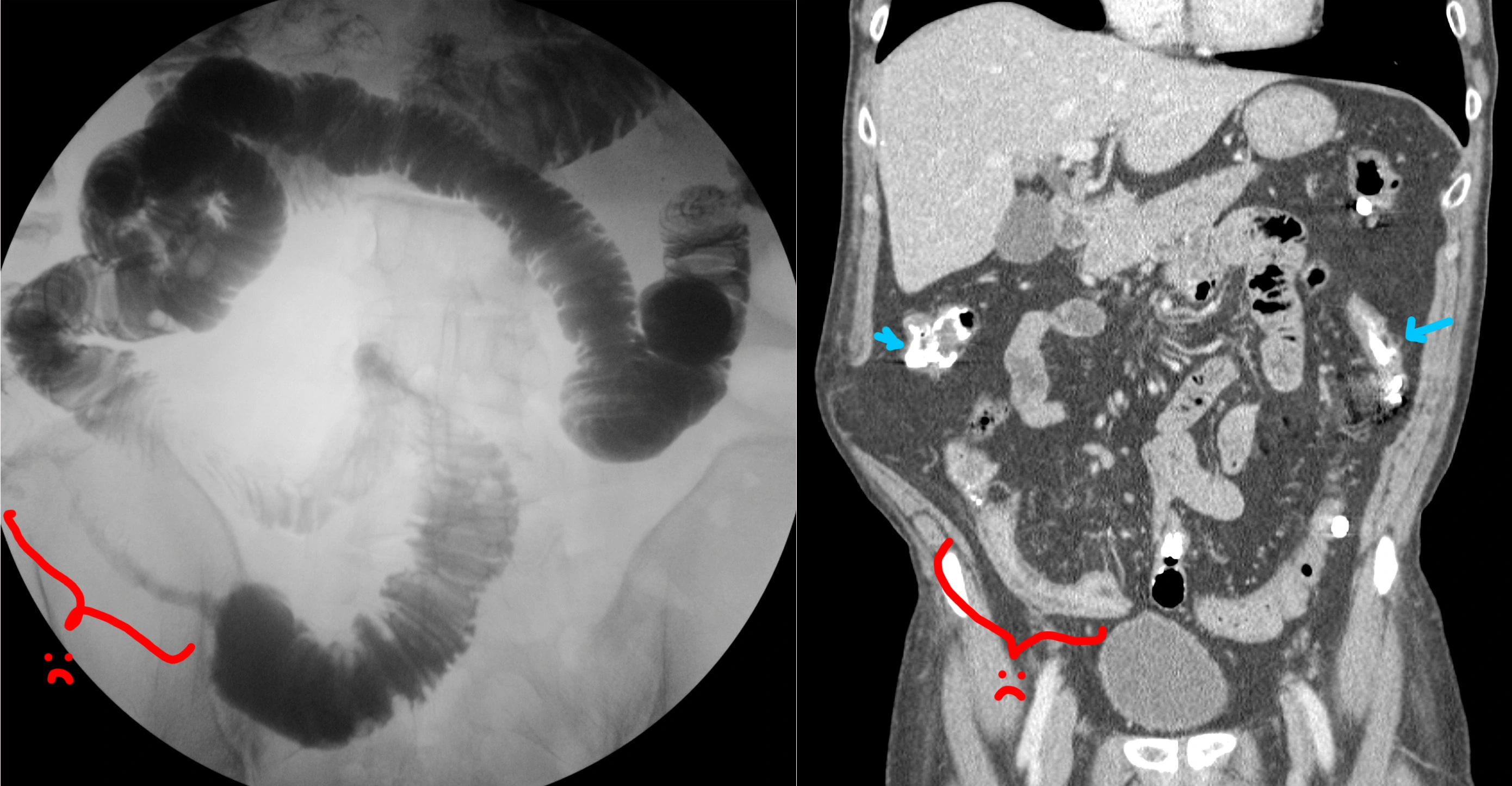
No clinical history saved on this one - sorry.
[Right] Small bowel follow-through (SBFT), where the patient drinks barium, and then we wait a bit until that barium is in the small bowel, then we take some pictures. This study is showing a long segment of terminal ileum that is strictured and severely narrowed in fibrostenotic Crohn's (red bracket). This is called the "string sign."
[Left] Coronal CT performed sometime after the SBFT. You can still see some residual barium in the small and large bowels (blue arrows). Red bracket shows the CT appearance of the terminal ileum stricture. On the CT, you can also see that the strictured segment has submucosal fat deposition, the "fat halo sign."
Two patients showing the varied appearance of large B-cell lymphoma of the stomach.
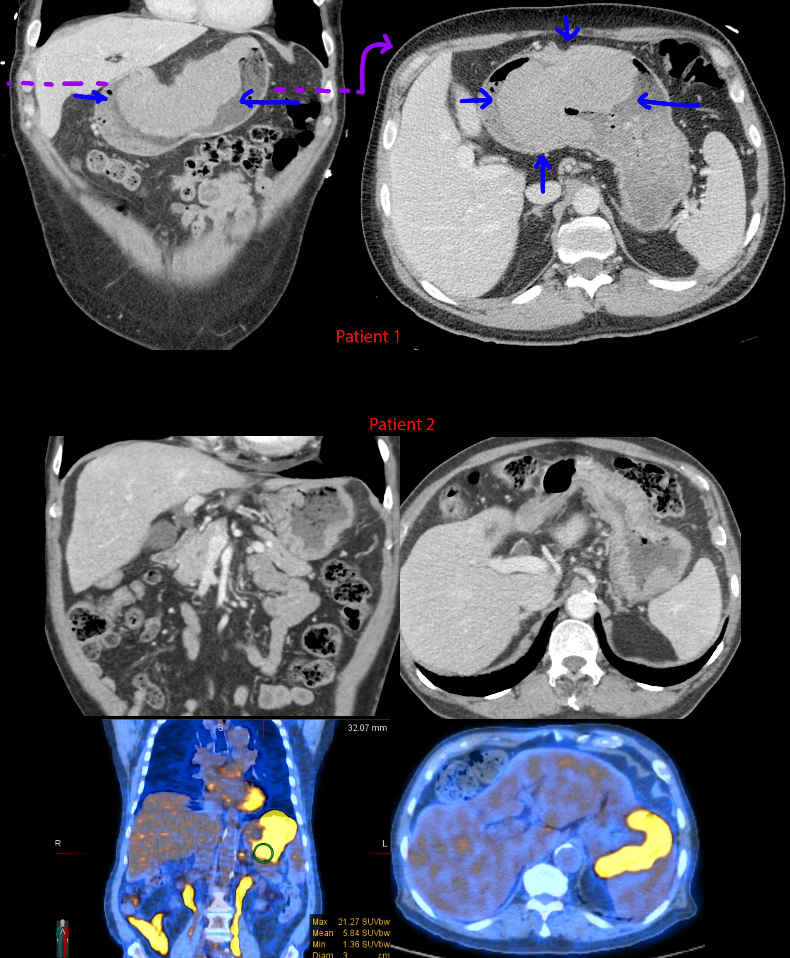
I didn't save any clinical history for these - sorry.
[Top] Patient 1 - Gigantic mass along the lesser curvature of the stomach. Look down at your belly - this mass is about 1/3rd the width from left to right.
[Mid] Patient 2 - CTs showing gently lobulated and undulating wall thickening of the gastric cardiac and fundus. Notice the transition from the normal gastric rugae to the smoother wall thickening where it is infiltrated by lymphoma. There is also mild (aneurysmal) dilation of the stomach where the wall thickening is located.
[Bottom] Patient 2 - PET-CT. The wall thickening is ridiculously hypermetabolic with a max SUV of 21.3. For comparison, the liver is normally in the range of 2-4 mean SUV.
Tuberculosis, sarcoidosis, lymphoma, and metastatic disease - these 4 can look like almost anything.
Psychiatric patient was eating a lot of hair (trichobezoar, gastric perforation).

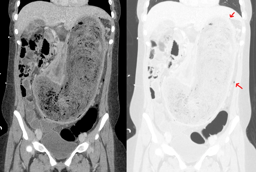
This patient with a long psychiatric history was found to be eating a lot of hair and eventually starting vomiting.
CT shows a large mass completely distending the stomach consistent with a giant hairball based on the given history.
CT in the lung/air window shows a few tiny foci of air outside of the stomach (arrows), not where there should be any air at all. This is consistent with a small perforation of the stomach.
See here for another case of bezoar causing stomach obstruction.
Stomach cancer causing obstruction.

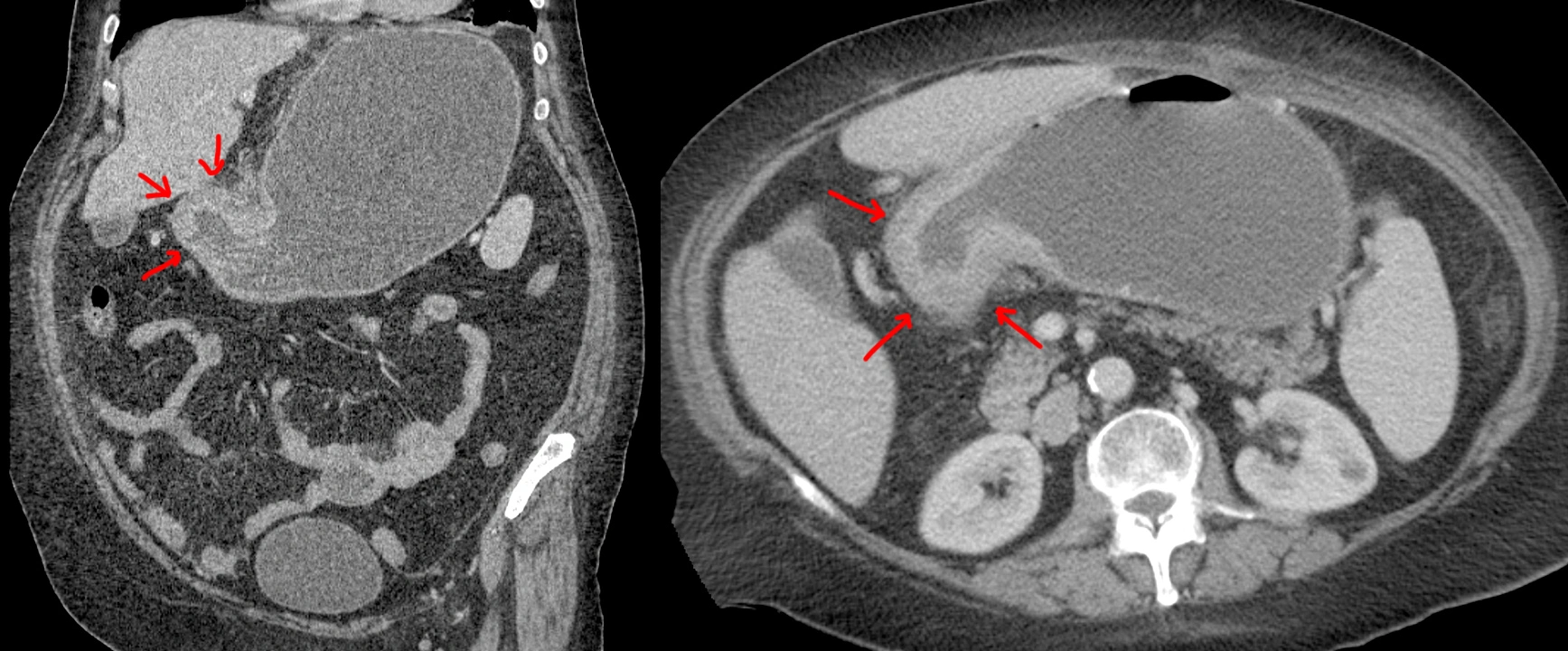
68 year old with nausea and vomiting.
CT shows very thickened irregular walls at the gastric outlet (arrows) with distended stomach.
Biopsy showed poorly-differentiated adenocarcinoma with signet ring cells.