Search
Cancer versus not cancer on esophagram.


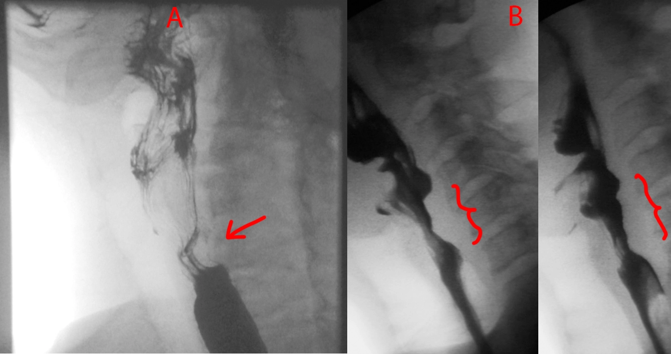
Quick one today. Take a look at Patient A and Patient B.
Patient A has a smooth focal indentation of the posterior cervical esophagus.
Patient B has a broader indentation that is also irregular and nodular along its contour.
Patient A has a cricopharyngeal bar, which is a prominence caused by the cricopharyngeus muscle that can cause dysphagia if it gets really prominent. Patient B has esophageal squamous cell carcinoma.
Conjoined twin, from prenatal to postnatal.


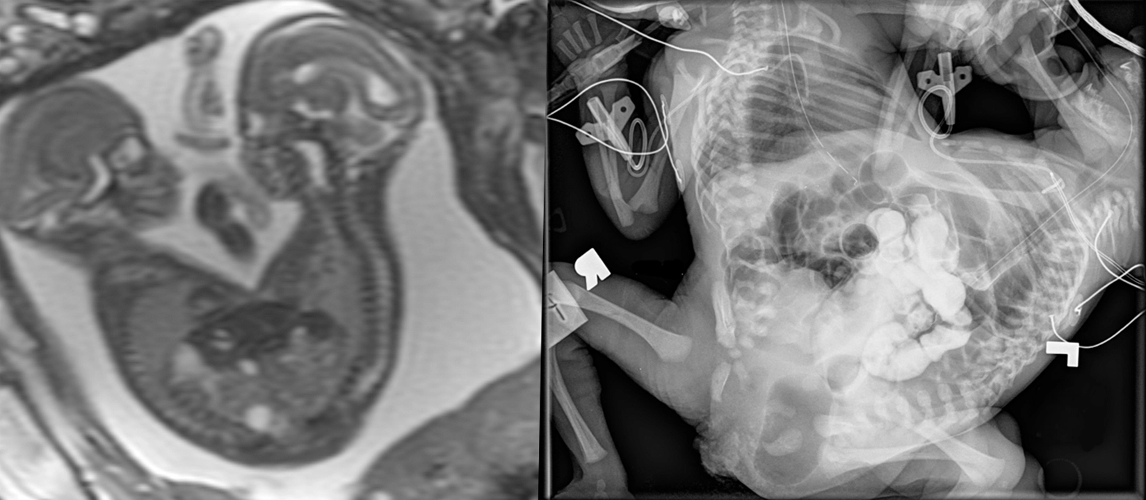
[Left]: Fetal MRI (FIESTA sequence) shows twins joined from their lower chest to the pelvis, but truly fused and sharing a single abnormal pelvic region. Not shown, but there are 3 lower limbs - one of the twins only had a single lower extremity.
[Right]: Postnatal small bowel follow-through (SBFT). It was unclear initially whether the twins shared a single rectum or had their own rectum. Therefore, contrast was administered via nasogastric tube for the twin with the suspected nonfunctional rectum, and serial imaging was performed until it passed into what turned out to be a separate, functional, but small rectum/anus.
I do not know too much about conjoined twins - not my area of expertise, but the general forms to consider are the side of fusion: ventral (front to front), lateral (side to side), dorsal (back to back), or caudal (tail end to tail end). Within these first 3, there are subtypes depending on how far up the fusion goes (head, chest, abdomen/pelvis); by definition, the caudal version obviously is only a lower body fusion. Once this is derived, an additional classification is the number of upper and lower limbs.
Crohn's disease.


No clinical history saved on this one - sorry.
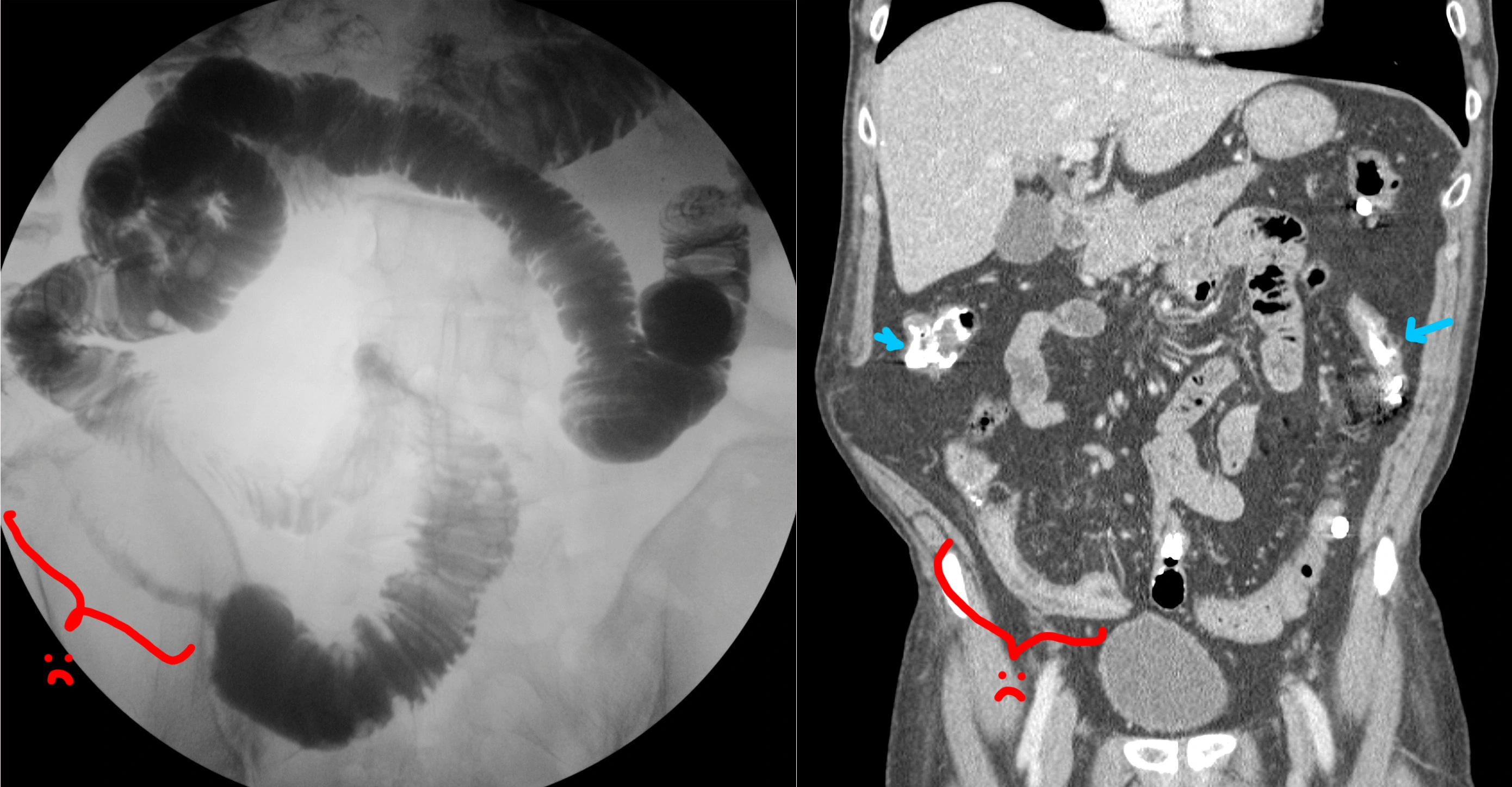
[Right] Small bowel follow-through (SBFT), where the patient drinks barium, and then we wait a bit until that barium is in the small bowel, then we take some pictures. This study is showing a long segment of terminal ileum that is strictured and severely narrowed in fibrostenotic Crohn's (red bracket). This is called the "string sign."
[Left] Coronal CT performed sometime after the SBFT. You can still see some residual barium in the small and large bowels (blue arrows). Red bracket shows the CT appearance of the terminal ileum stricture. On the CT, you can also see that the strictured segment has submucosal fat deposition, the "fat halo sign."