Search
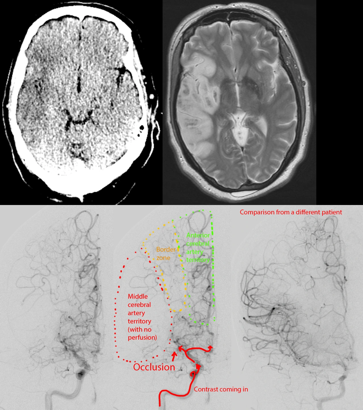
Large acute stroke.


Female in her 50s, with a history of meth abuse. She suddenly developed confusion and left-sided weakness while talking on the phone.
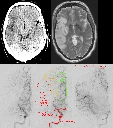
Dual energy CT virtual noncontrast [top left image] shows a large rectangular area of hypodensity in the right middle cerebral artery territory.
Cerebral angiogram shows occlusion of the right MCA with no filling of its distal branches or the brain parenchyma [bottom left, bottom middle]. A stent retriever mechanical thrombectomy was performed [not shown].
MRI done 1 day later shows the infarct with the affected area showing brain swelling [top right].
Next